Malaria Cell Images



Overview
The NIH Malaria Cell Images dataset contains 27,558 segmented red blood cell crops from Giemsa-stained thin blood smears. The task is binary classification of parasitized versus uninfected cells, not patient-level diagnosis.
Train: 27,558 images, with 13,779 per class
Images: PNG crops with variable dimensions, converted to RGB without resizing
Official splits: none; the builder exposes the entire release as
trainDownloads: the cell-image archive and two NIH patient-to-cell mapping CSVs, each verified against a pinned SHA-256 checksum
License: not stated on the NIH datasheet; cite the original paper when using the data
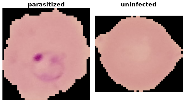
Data Structure
Accessing ds[i] returns a dictionary with these fields:
Key |
Type |
Description |
|---|---|---|
|
|
RGB red blood cell crop with its original dimensions |
|
int |
Infection class index (0 or 1) |
|
str |
Original cell-crop filename, including the |
|
str |
Identifier from the class-specific NIH patient-to-cell CSV, with surrounding whitespace stripped |
|
str |
Microscopy image identifier: the filename prefix before |
Raw access with ds.with_format("raw") returns encoded image bytes instead
of decoded PIL images. Examples are ordered deterministically by archive path.
The supervised keys are ("image", "label"). The builder skips non-image
archive entries such as Thumbs.db and raises an error if an accepted cell
image has no patient-mapping entry or cannot be decoded.
Classes
ds.features["label"].names lists class names in this fixed index order:
Label |
Class Name |
Images |
|---|---|---|
0 |
|
13,779 |
1 |
|
13,779 |
Downloads and Checksum Provenance
Malaria.SOURCE.assets declares exactly three downloads:
cell_images.zip: the single-cell classification archive, containing thecell_images/Parasitizedandcell_images/Uninfecteddirectories. Its pinned SHA-256 digest comes from the TensorFlow Datasets Malaria checksum manifest linked below.patientid_cellmapping_parasitized.csv: the NIH patient-to-cell mapping used to populatepatient_idfor parasitized cells.patientid_cellmapping_uninfected.csv: the corresponding mapping for uninfected cells.
The two CSV SHA-256 digests were computed locally from the official NIH downloads and pinned in the builder. They detect changes relative to those downloaded versions; they are not publisher-provided checksum attestations. All three downloads are checked against the builder’s pinned digests when newly downloaded. The CSVs are auxiliary metadata, not additional dataset splits.
The other datasets listed on the NIH datasheet, including full-resolution smear images, thick smears, and Vivax datasets, are separate releases and are not downloaded by this builder.
Usage Example
from stable_datasets.images import Malaria

# Downloads and prepares the release on first use; subsequent loads reuse the cache.
ds = Malaria(split="train")
print(len(ds))  # 27558
sample = ds[0]
class_names = ds.features["label"].names
print(class_names[sample["label"]])
print(sample["patient_id"], sample["source_image_id"])

# Omitting split returns a StableDatasetDict containing only train.
ds_all = Malaria()
print(list(ds_all))  # ['train']
Requesting test or validation raises an error: those splits are not
published, and the builder does not invent them.
Leakage-Aware Splitting
Cells from the same patient can share staining, acquisition, and microscopy
characteristics. A random cell-level split can place a patient’s crops in both
training and validation, producing optimistic results. Group by the provided
patient_id when constructing a patient-disjoint evaluation. Grouping only
by source_image_id is weaker because a patient can contribute multiple images.
For example, hold out 20% of the patient identifiers using GroupShuffleSplit:
from sklearn.model_selection import GroupShuffleSplit
from stable_datasets.images import Malaria

ds = Malaria(split="train")
# Read metadata without decoding every image.
patient_ids = [row["patient_id"] for row in ds.with_format("raw")]
splitter = GroupShuffleSplit(n_splits=1, test_size=0.2, random_state=42)
train_indices, validation_indices = next(
    splitter.split(range(len(ds)), groups=patient_ids)
)
train_ds = ds.select(train_indices)
validation_ds = ds.select(validation_indices)

train_patients = {patient_ids[index] for index in train_indices}
validation_patients = {patient_ids[index] for index in validation_indices}
assert train_patients.isdisjoint(validation_patients)
The hold-out fraction refers to patient groups, not cells, and does not
guarantee class balance. Inspect per-class counts after splitting. These are
user-created subsets of the published train release, not official splits.
The benchmark harness’s default random 10% hold-out (seed 42) is a cell-level
evaluation and should not be presented as patient-disjoint performance.
References
Kather Colorectal Histology: a complementary multi-class histology benchmark
Citation
@article{rajaraman2018pre,
  title={Pre-trained convolutional neural networks as feature extractors toward
         improved malaria parasite detection in thin blood smear images},
  author={Rajaraman, Sivaramakrishnan and Antani, Sameer K and Poostchi, Mahdieh
          and Silamut, Kamolrat and Hossain, Md A and Maude, Richard J and Jaeger,
          Stefan and Thoma, George R},
  journal={PeerJ},
  volume={6},
  pages={e4568},
  year={2018},
  doi={10.7717/peerj.4568}
}